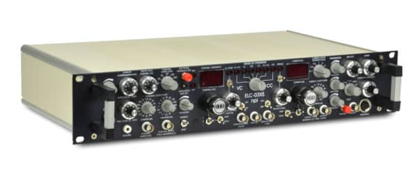
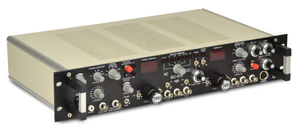
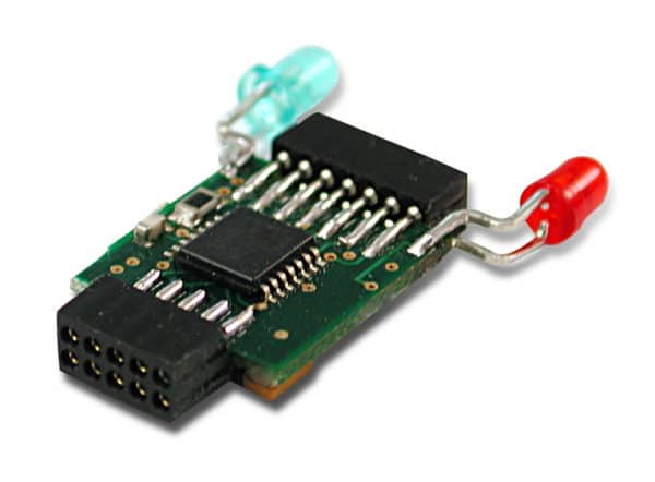
The ELC electrophysiology amplifiers are wide-bandwidth high impedance universal amplifiers. The versatile design allows intracellular and extracellular recordings as well as current or voltage application, all with the same headstage. It can be used with sharp microelectrodes, patch clamp pipettes, perforated or tight seal patch configurations and features electroporation and iontophoresis functionalities. It is also possible to perform extracellular recordings due to versatile high- and lowpass Bessel filters.
npi’s ELC electrophysiology amplifiers are used in vertebrate (rat, mouse, cat, fish etc.) and invertebrate (leech, crab, snail, insect etc.) neurons, muscle cells, other excitable tissue and plant cells. They can be used in-vitro and in-vivo and have been referenced in numerous publications.
Check out our tutorial videos about the ELC-03XS Universal Amplifier on YouTube


The ELC universal electrophysiology amplifier series can be used for:
• Whole cell patch clamp recordings
• Sharp microelectrode intracellular recordings
• Extracellular recordings
• Juxtacellular recordings
• Juxtacellular filling
• Single cell electroporation
• Single cell stimulation
• Iontophoresis
• Voltammetric recordings
The single-cell juxtacellular recording–labeling technique makes it possible to label the neuron recorded extracellularly. It is a very useful tool for achieving single-cell structure–function correlation studies in living, intact neural networks and for determining their phenotype and genotype. It can reveal the overall picture of the smallest neurons, including interneurons. It can be combined with other electrophysiological techniques (e.g. electro-encephalographic recordings and intracerebral electrical stimulation), electron microscope, immunohistochemical, molecular and/or genetic techniques.
Its principle consists in iontophoresing tracer molecules into the membrane of the neuron being recorded. This is done under continuous electrophysiological monitoring, allowing the retrieval of the neuron labeled in more than 85% of attempts. Continuous DC recordings suggest that the juxtacellular filling “or tickling” procedure produces a transient micro-electroporation, which allows the internalization of the tracer into the intracellular space. Since this procedure allows single neurons to be recorded for long periods, many electrophysiological features can be collected, and the finest and remotest axonal ramifications can be marked. In spite of some limitations and pitfalls, the juxtacellular technique remains the high standard for investigating the genetic, molecular, physiological, and architectural bases of cell–cell communication. It is also a very versatile and useful tool when it comes to decipher, for instance, the molecular, cellular, and network mechanisms of brain state, physiological, and pathological oscillations.
The ELC electrophysiology amplifiers are well suited for precise voltage clamp, for whole cell patch clamp, loose patch clamp or cell attached patch clamp.
Yes, you can. It is easy to use in head-fixed animals. npi also provides miniature headstages for current clamp recordings in freely moving animals.
There is a red pushbutton named BUZZ on the front panel. As soon as this is pushed, the amplifier’s capacity compensation goes to maximum, which results in rapid oscillations in the headstage’s output current. This effect, which is unwanted during regular recordings leads to a very localized mechanical stress to the cell’s membrane – small holes are generated and the electrode tip can impale into the cell.
Our dedicated team has years of industry and laboratory experience.
From complex technical challenges to tailored product recommendations,
our application scientists are ready to support your needs with hands-on expertise.